
- Researchers have created the most intricate functional brain map, focusing on a mouse brain segment with 84,000 neurons and 500 million synapses.
- This groundbreaking study involved capturing neuronal activity while a mouse watched various films, using advanced imaging techniques.
- The visual cortex was dissected into 25,000 ultra-thin layers, generating nearly 100 million high-resolution images to construct a 3D brain map.
- Artificial intelligence helped depict the complex neural circuitry, unveiling communication pathways more intricate than a city skyline.
- This endeavor parallels the Human Genome Project, offering insights for future research on disorders like Alzheimer’s and autism.
- The public release of the brain map invites global collaboration and exploration of new hypotheses and therapies.
- The MICrONS consortium aims to further decode the entire mouse brain, expanding our understanding of brain complexity.
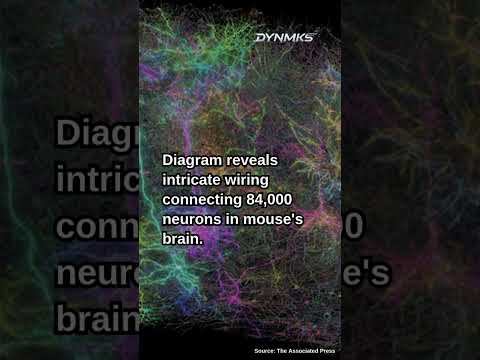
In a groundbreaking convergence of science fiction and cutting-edge neuroscience, researchers have accomplished a herculean task: crafting the most intricate functional brain map ever forged. The star of this neural odyssey, a small segment of a mouse brain, was showcased like never before, revealing the vast labyrinth of cerebral connections that define cognition and behavior. Visualize this: 84,000 neurons linked by a staggering 500 million synapses, all housed in a fragment no larger than a poppy seed. This monumental achievement stands as a testament to human ingenuity and curiosity.
The journey began at Baylor College of Medicine, where a genetically engineered mouse was shown a medley of sci-fi clips, sports, and nature scenes. As the minutes of film unfolded, a laser-powered microscope danced along, capturing the vivid activity within the mouse’s visual cortex. The mouse became an unwitting luminary in this visual symphony, its neurons flickering away under the gaze of sophisticated technology.
The visual cortex, vibrant and active, was taken to the Allen Institute, where it underwent a transformation akin to slicing a grand tome into ultra-thin pages—25,000 layers deep. From these slivers emerged nearly 100 million high-resolution electron microscope images, the foundation for a vivid three-dimensional map of the brain’s intricate circuitry.
In this map, each neural pathway unfurled like a richly hued tapestry, its complexity captured by artificial intelligence at Princeton. The interconnected synapses, each distinguished by individual colors, painted a picture of communication more intricate than any Manhattan skyline. With the neural wiring traced akin to a map stretching three miles if unraveled, a new understanding of how visual input flows through the brain took shape.
To many scientists, this endeavor mirrors the ambition and impact of the Human Genome Project. As they gaze upon these cerebral vistas, researchers are plotting future routes toward demystifying disorders such as Alzheimer’s and autism. This venture, shouldered by 150 researchers from various corners of the globe, gives rise to limitless possibilities. The brain map’s public release invites not only seasoned scientists but also inquisitive minds everywhere to delve into a hitherto unseen world, promising new hypotheses and therapeutic avenues.
The study reminds us of the brain’s unparalleled complexity and inspires a sense of discovery akin to peering into a universe—both astronomical and neuron-sized. The MICrONS consortium, buoyed by this triumph, looks ahead with anticipation, setting sights on an even bolder goal: to decode an entire mouse brain.
Amidst the sparks of neuron-generated luminescence, a clear message resounds: the journey to understanding our own minds is as boundless as space itself, one mouse-brain movie marathon at a time.
Inside the Neuron Network: A New Era in Brain Mapping
The recent feat of mapping an intricate segment of a mouse brain has captured widespread attention in the scientific community. Designed by a global team of researchers, this pioneering project opens new avenues not only for neuroscience but also for practical applications across diverse fields.
How the Marvel Was Achieved
Advanced Imaging Techniques: Utilizing a laser-powered microscope, researchers captured the neuronal activity in a mouse’s visual cortex at Baylor College of Medicine. This technique allowed them to trace vibrant neural communication pathways as the mouse watched various film clips.
High-Resolution Imaging: At the Allen Institute, the mouse’s brain was sliced into 25,000 ultra-thin layers, resulting in 100 million high-resolution images. These formed the basis for a three-dimensional map, comparable to turning an intricate brain into a navigable city modeled by AI at Princeton.
Artificial Intelligence in Brain Mapping: AI was pivotal in deciphering the complex neuron interactions. By highlighting different synapses with unique colors, AI helped visualize the interwoven communication networks that teem within even the smallest brain segments.
Unexplored Synergies and Applications
Real-World Use Cases:
– Medical Research: This map provides a foundational tool for understanding neural pathologies associated with disorders like Alzheimer’s and autism. Insights from this study could lead to predictive models or new treatment strategies.
– Enhanced Brain-Computer Interfaces: Insights into neural communication could enhance the development of brain-controlled prostheses and advanced AI systems that mimic cognitive processes.
Industry Trends and Forecasts:
– Biotechnology Growth: Projects like this could drive growth in biotechnology, especially in neuroimaging and computational neuroscience.
– AI Integration in Neurosciences: The successful application of AI here signals an increasing trend towards AI-driven neuroscience research, likely a major driver in future breakthroughs.
Pressing Questions
Why Study a Mouse Brain?:
– Mouse brains share many anatomical and functional similarities with human brains, making them an excellent model for understanding human neurology.
How Will This Impact Daily Life?:
– Long-term, this research could lead to more effective neurological and psychological therapies, improving quality of life for millions.
Pros & Cons Overview
Pros:
– Detailed Insight: Unprecedented detail in capturing brain functions offers immense research potential.
– Broadened Collaboration: The public release of this data invites collaboration and discovery from a wider scientific community.
Cons:
– Scale Limitation: The study only covers a small segment of the mouse brain; scaling this to larger systems poses technical challenges.
– Resource Intensive: The process is costly and time-consuming, which could delay broader applications.
Actionable Recommendations
– For Researchers: Dive into the publicly available dataset to explore new research questions or validate previous hypotheses.
– For Educators: Use this brain map as a teaching tool to inspire interest in neuroscience and data science.
– For Entrepreneurs: Monitor advances in brain mapping and AI to identify new business opportunities in therapy and technology solutions.
For more cutting-edge insights, visit the Allen Institute.
This monumental achievement marks a key moment in neuroscience, heralding a future where the enigmatic brain may become far more intelligible, ushering in a new era of scientific discovery and innovation.


